Ear Anatomy
To better understand the cause of your hearing loss, you must first understand how the ear is designed and the conditions that interfere with hearing.
Sound travels through the air as a sound wave. Your ear turns the sound waves into information which your brain hears as sound.
First, the sound waves are collected by the pinna (outer ear) and travel down the canal to the eardrum. The pressure of the sound waves on the eardrum causes the eardrum to vibrate. There are three ossicles (tiny bones) in the middle space, behind the eardrum. When sounds vibrate the eardrum, the ossicles are set into motion causing the cochlea (fluid in the inner section) to move. The movement of fluid in the cochlea causes tiny hair cells to bend, sending electrical impulses through the auditory (hearing) nerve up to the brain where these electrical impulses are perceived as sound.
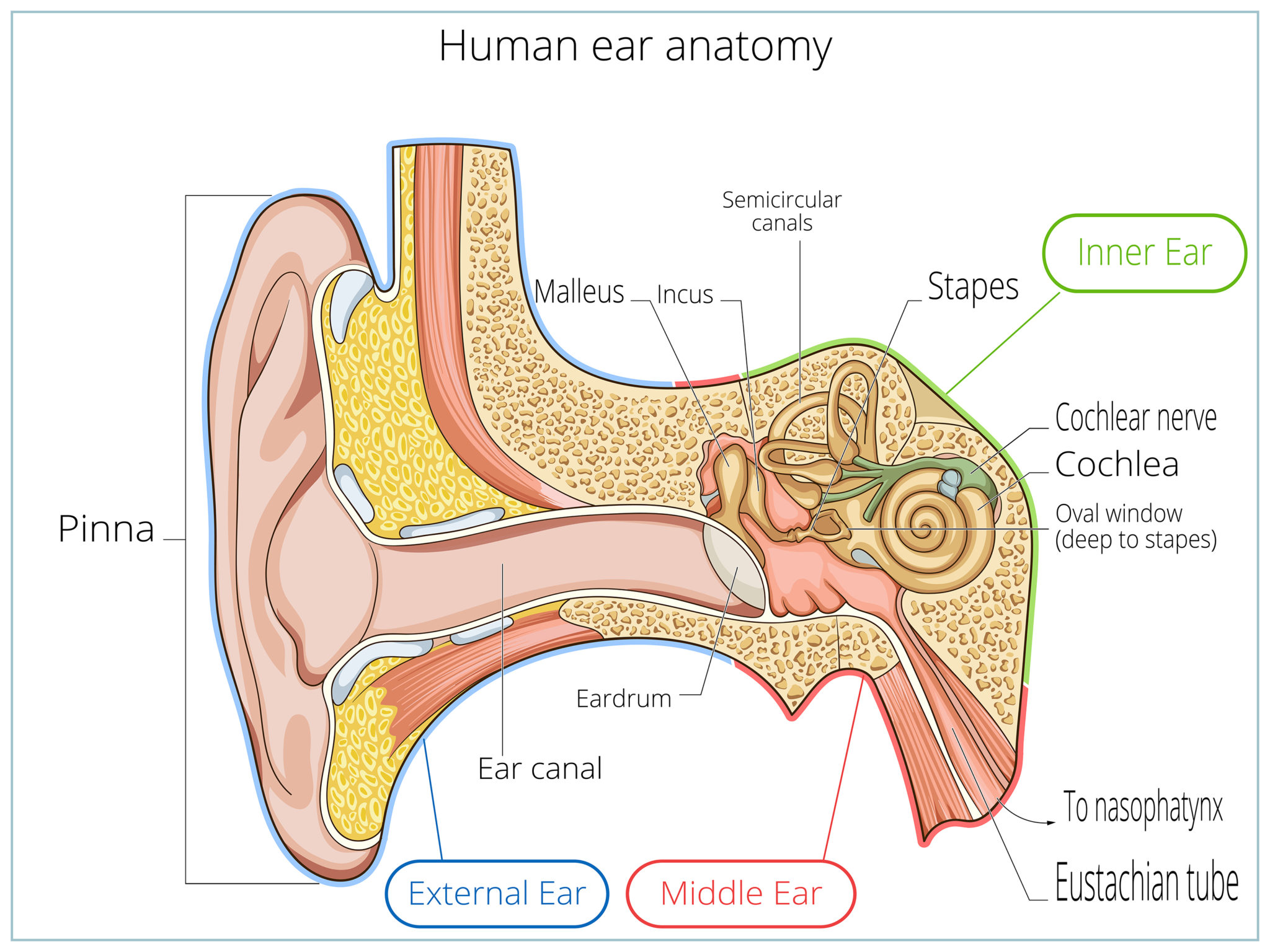
The Outer Ear
Consists of the visible part, called the pinna, and the canal.
The pinna collects and funnels the sound down the canal to the tympanic membrane (eardrum). The canal is made up of only a few layers of skin and small hairs. It has small glands that produce cerumen (wax) to help lubricate and protect the skin in the canal.

The Middle Ear
The tympanic membrane, or eardrum, divides the outer and middle ear.
The middle ear is an air-filled cavity that is also connected to the back of the nose through the eustachian tube. There are three small bones in the middle called ossicles. These tiny bones are called the malleus, incus, and stapes and they form a connected chain in the cavity from the eardrum to the inner section. The ossicles relay mechanical vibrations that are received at the tympanic membrane to the oval window which is the beginning of the cochlea.

The Inner Ear
Consists of two main structures
1) The semicircular canals
2) The cochlea
1) The semicircular canals do not contribute to hearing. However, they are important for balance.
2) The cochlea is the organ of hearing in the inner ear. The cochlea changes the mechanical vibrations from the middle section into electrical impulses in the inner section. The cochlea is a fluid-filled structure that looks like a snail. When the ossicles relay mechanical vibrations to the inner ear, the fluid in the cochlea begins to move. Small cells, called hair cells, bend in response to the movement of the fluid causing electrical impulses to be sent to the brain through the auditory nerve where it will be interpreted as sound.
Ready to learn more about the signs?
The signs of hearing loss usually emerge slowly over time. Learn more about common indications of hearing loss.